
02 SURFACE MARKINGS
Let's get right into it! Practice identifying the surface markings on your friends, or drawing them out.

What are the surface markings for the facial artery pulse?
- ((Posterior border of SCM::Incorrect))
- ((Anterior border of masseter::☑️ The facial artery can be palpated where it crosses the anterior border of the masseter muscle))
- ((Angle of the mandible posteriorly::Not the pulse point))
- ((Over the zygomatic arch::Superficial temporal artery))
What are the surface markings for the mental nerve?
- ((Below the canine tooth::Too medial))
- ((In line with the second premolar tooth::☑️ The mental foramen lies in line with the second premolar))
- ((At the angle of the mandible::Incorrect))
- ((At the midpoint of the chin::Incorrect))
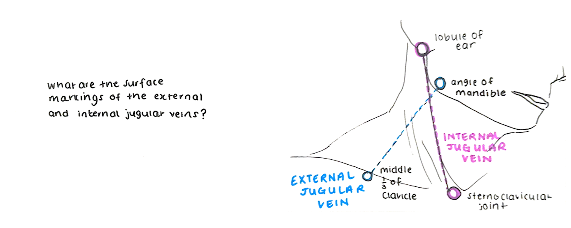
What are the surface markings for the internal jugular vein?
- ((Styloid process to medial end of clavicle::Styloid process is too deep; not a surface landmark))
- ((Ear lobule to medial end of clavicle::☑️ The internal jugular vein runs from the lobule of the ear to the sternoclavicular joint))
- ((Mastoid process to medial end of clavicle::Closer to the course of the SCM attachment, not the standard surface marking))
- ((Lesser cornu of hyoid to medial end of clavicle::Hyoid is not used as the superior surface landmark))
What are the surface markings for the external jugular vein?
- ((Lobule of ear to sternoclavicular joint::This describes the internal jugular vein))
- ((Angle of mandible to middle third of clavicle::☑️ The external jugular vein runs superficially from the angle of the mandible to the midpoint of the clavicle))
- ((Mastoid process to medial end of clavicle::Too medial; not the standard surface marking))
- ((Hyoid bone to lateral end of clavicle::Incorrect superior landmark))
A child inhales a peanut. Where is it most likely to lodge?
- ((Left lower lobe bronchus::The left main bronchus is narrower and more horizontal))
- ((Left upper lobe bronchus::Less common site))
- ((Right lower lobe bronchus::☑️ The right main bronchus is wider, shorter, and more vertical, so foreign bodies most commonly pass into the right lower lobe bronchus))
- ((Right upper lobe bronchus::Less common than lower lobe))
👩⚕️ The right main bronchus is wider, shorter, and more vertical than the left, so foreign bodies preferentially enter it, especially into the lower lobe bronchus.
Where is the duodenojejunal (DJ) flexure located under normal anatomical conditions?
- ((Right of the midline at L2::Right-sided DJ flexure suggests malrotation))
- ((Left of the midline at the transpyloric plane (L1)::☑️ Normal position — to the left of the spine at the level of the transpyloric plane))
- ((Midline at L3::Incorrect vertebral level))
- ((Right upper quadrant below the liver::Abnormal position))
👩⚕️ Malposition of the DJ flexure to the right is a sign of malrotation!
What are the surface markings for the gallbladder fundus?
- ((Tip of the right 7th costal cartilage::Too superior))
- ((Tip of the right 9th costal cartilage in the midclavicular line::☑️ The fundus of the gallbladder lies at the tip of the right 9th costal cartilage)))
- ((McBurney's point::Appendix landmark))
- ((Left 9th costal cartilage::Incorrect side))
What are the surface markings of McBurney’s point?
- ((One third from the ASIS to the umbilicus::☑️ McBurney’s point lies at the junction of the lateral one-third and medial two-thirds of the line from ASIS to umbilicus))
- ((Midpoint between the ASIS and pubic symphysis::This is the mid-inguinal point, not McBurney’s point))
- ((Midpoint between the ASIS and pubic tubercle::This is the midpoint of the inguinal ligament))
- ((Two thirds from the ASIS to the umbilicus::Incorrect position))
What are the surface markings for femoral artery access?
- ((Midpoint between ASIS and pubic symphysis::☑️ The femoral artery lies at the mid-inguinal point, between the ASIS and pubic symphysis))
- ((Midpoint between ASIS and pubic tubercle::This is the midpoint of the inguinal ligament, not the femoral artery location))
- ((Medial to the pubic tubercle::The femoral artery lies at the mid-inguinal point, between the ASIS and pubic symphysis))
- ((Lateral to the ASIS::Not anatomically related to the femoral artery))
👩⚕️ The mid-inguinal point is the midpoint between the ASIS and pubic symphysis and is the surface landmark for the femoral artery. The midpoint of the inguinal ligament is the midpoint between the ASIS and pubic tubercle and is the surface landmark for the deep inguinal ring.